Bubble Study (Contrast Echo)
An echocardiogram shows the anatomy, structure and function of your heart.
A common type of echocardiogram is called a transthoracic echocardiogram. In this test, sound waves directed at your heart from a wand-like device (transducer) held on your chest produce video images of your heart in motion. Doctors may use this test to diagnose a patent foramen ovale and detect other heart problems.
Variations Of This Procedure May Be Used To Identify Patent Foramen Ovale, Including:
• Colour flow Doppler. When sound waves bounce off blood cells moving through your heart, they change pitch. These characteristic changes (Doppler signals) and computerized colourization of these signals can help your doctor examine the speed and direction of blood flow in your heart.
• If you have a patent foramen ovale, a colour flow Doppler echocardiogram could detect the flow of blood between the right atrium and left atrium.
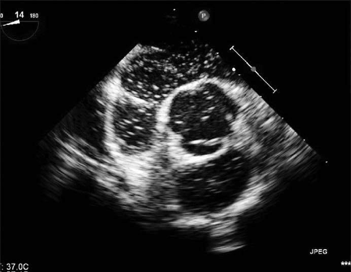
• Saline contrast study (bubble study). With this approach, a sterile salt solution is shaken until tiny bubbles form and then is injected into a vein. The bubbles travel to the right side of your heart and appear on the echocardiogram.
If there's no hole between the left atrium and right atrium, the bubbles will simply be filtered out in the lungs. If you have a patent foramen ovale, some bubbles will appear on the left side of the heart. The presence of a patent foramen ovale may be difficult to confirm by a transthoracic echocardiogram.